Dr. Nilkanth C. Patil is a renowned Interventional Cardiologist in Hyderabad, known for his expertise in complex heart procedures and advanced cardiac imaging. As a highly respected cardiologist and heart specialist, he brings 26 years of overall medical experience, with 15 years dedicated to cardiology. His clinical journey includes advanced training from premier institutes in India and abroad.
After completing his MBBS from LTMMC, Mumbai, Dr. Patil pursued an MD in Internal Medicine from BMCRI, Bangalore. He earned his DM in Cardiology from AIIMS, New Delhi, securing the top rank in the national entrance exam. His dedication to clinical excellence led him to gain international exposure through a fellowship at the London Health Sciences Centre in Ontario, Canada. Throughout his career, he has held senior positions in reputed hospitals, including Metro Hospital (Delhi), Care Hospitals (Hyderabad), and AIG Hospitals, where he is currently practicing.
Dr. Patil’s areas of special interest include coronary imaging and interventions, congenital heart defect device closure, and structural heart interventions. He is also highly skilled in advanced 3D/4D echo, transesophageal echocardiography (TEE), cardiac CT, and MRI scan interpretation, making him a trusted choice for advanced and minimally invasive cardiac care.
- Call Us: 8885718260
- Email Us: drnilpatil@gmail.com
Leading Edge Heart Interventions
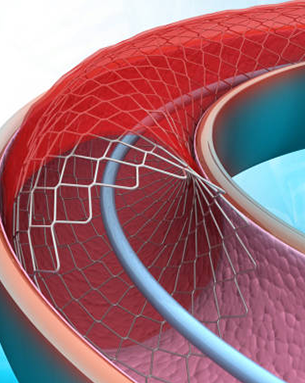
Primary Angioplasty with Surgical Precision
 (1).png)
Mastering Complex Coronary Cases
Advanced angioplasty for critical blockages

New Life for Failing Valves
Transcatheter Aortic Valve Replacement for faster recovery & function

.png)
.png)
.png)
Dr. Nilkanth C. Patil
About Us
Dr. Nilkanth C. Patil is a highly skilled Interventional Cardiologist with extensive expertise in diagnosing and treating complex heart conditions. Specializing in coronary interventions and structural heart interventions, he offers advanced solutions for patients with heart blockages, valve disorders, and congenital heart defects. His proficiency extends to congenital heart defect device closure, helping patients with complex congenital heart conditions achieve better quality of life.
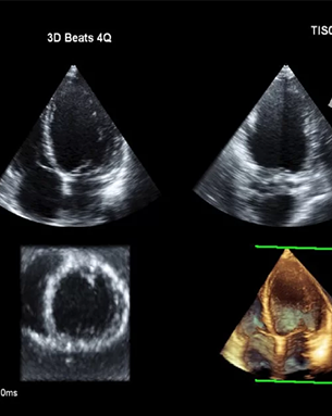
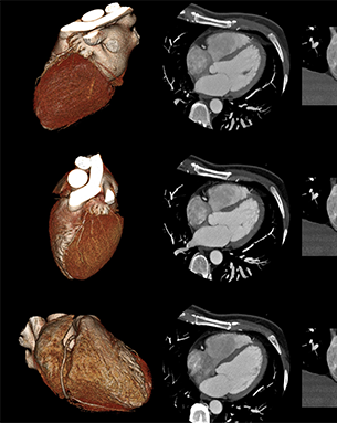
With advanced training in cardiac imaging, Dr. Patil employs the latest technologies like 3D/4D echo, transesophageal echocardiography (TEE), and cardiac CT and MRI scans to provide precise and comprehensive heart assessments. His deep understanding of heart anatomy and function allows him to tailor treatments that are minimally invasive and highly effective, ensuring optimal outcomes for his patients.
Dr. Patil is also committed to preventive cardiology, focusing on early detection and management of heart disease risks. Through personalized care plans, he empowers his patients to take charge of their heart health, minimizing the need for extensive interventions and promoting long-term wellness.
26+
Years of Overall Experience
15+
Years of Experience as Cardiologist
30K+
Happy Patients
Our Specialization

Coronary Interventions

Structural Heart Disease

Pulmonary Hypertension

Preventive Cardiology
Our Healthcare Services
Advanced Heart Imaging and Interventions


Dr. Nilkanth C. Patil
Interventional Cardiologist

- MBBS ( LTMMC, Mumbai ) MD Internal Medicine ( BMC, Bangalore ) DM Cardiology ( AIIMS, New Delhi) Fellowship in Cardiology in London Health Sciences Centre, London. Ontario. Canada
- 26.0 Years Of Experience
- Licence No. TSMC/FMR/30287
- Call
- Video Call
- In person
Testimonials
What our Patients says about us
Great experience with Dr. Nilkanth C. Patil. Accurate diagnosis, clear explanation, and effective treatment. Approachable and doesn't overprescribe. Thank you, Doctor.

Sunil
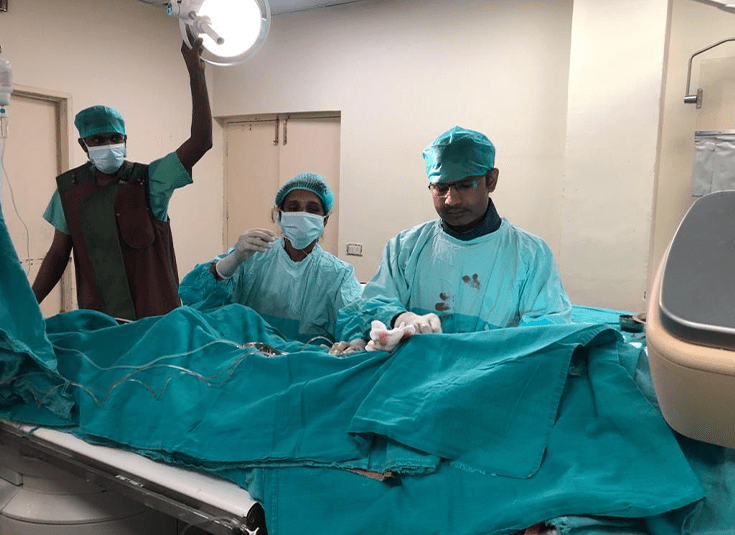
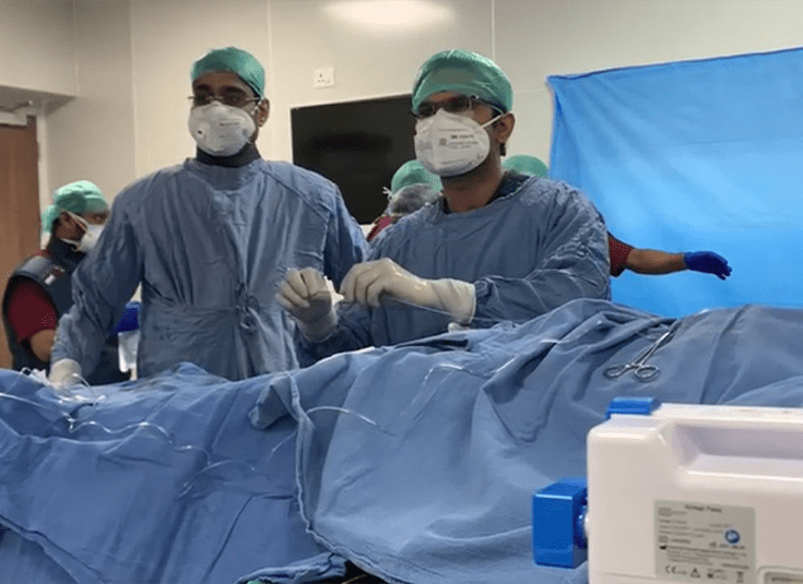
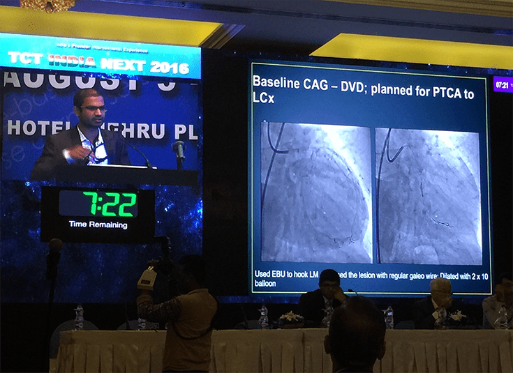
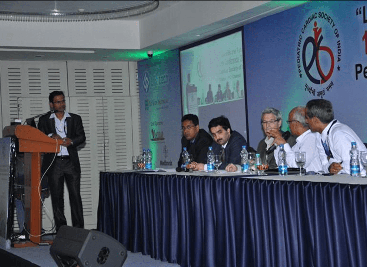
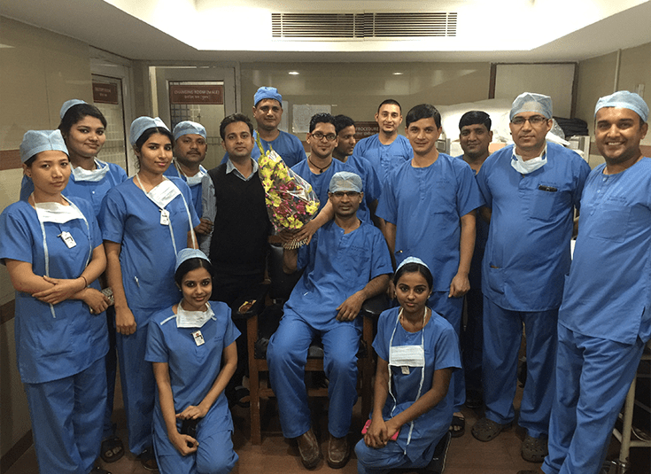
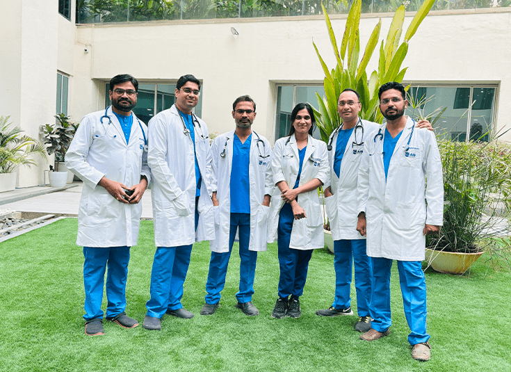
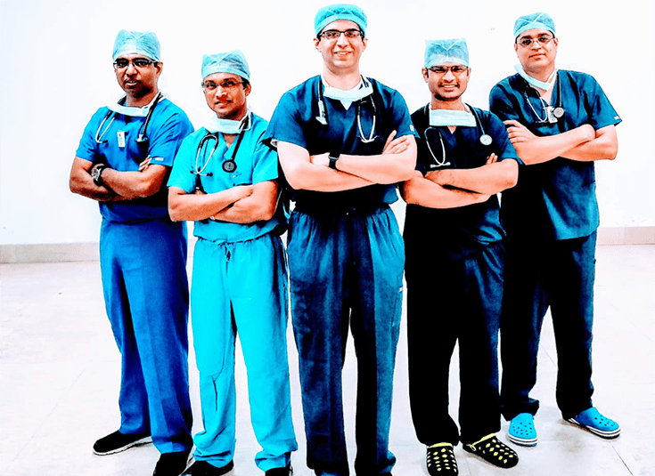
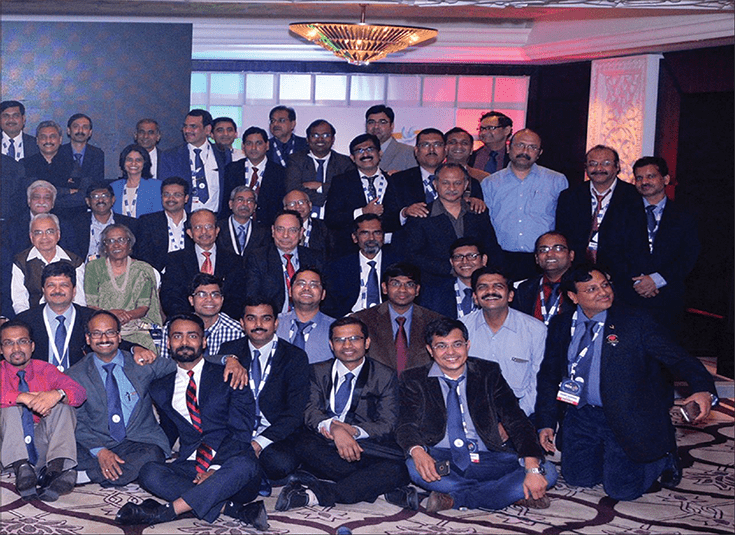
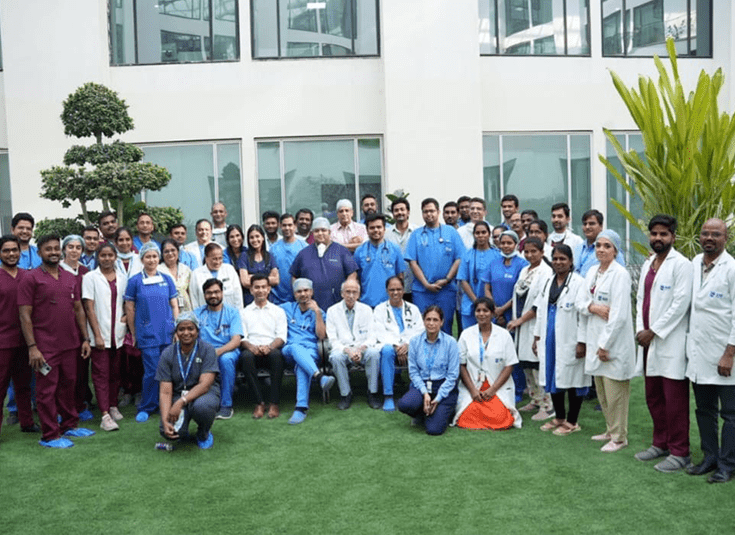
Media Pictures

Frequently Asked Questions
What sets Dr. Nilkanth C. Patil apart as an Interventional Cardiologist in Hyderabad?
Dr. Nilkanth C. Patil is a distinguished Interventional Cardiologist in Hyderabad, recognized for his 26 years of comprehensive medical experience, with 15 years specializing in cardiology. Trained at premier institutes like AIIMS, New Delhi, and with international fellowship at the London Health Sciences Centre, Canada, Dr. Patil excels in complex heart procedures and advanced cardiac imaging. His expertise in coronary imaging, structural heart interventions, and advanced 3D/4D echo makes him highly respected in his field.
What are Dr. Nilkanth C. Patil’s areas of expertise in heart care?
Dr. Nilkanth C. Patil, a renowned heart specialist in Hyderabad, specializes in a range of advanced cardiac procedures. His areas of expertise include coronary imaging and interventions, congenital heart defect device closure, and structural heart interventions. With a strong foundation in advanced 3D/4D echo, transesophageal echocardiography (TEE), cardiac CT, and MRI scan interpretation, Dr. Patil offers trusted, minimally invasive cardiac care. His seasoned experience at top hospitals enhances his reputation as a leading cardiologist.
What are coronary imaging and interventions?
Coronary imaging and interventions include techniques like angiography, which visualizes blood flow in coronary arteries. Advanced interventional procedures, like angioplasty and stenting, open blocked arteries to restore normal blood flow, preventing heart attacks. These procedures utilize advanced imaging for precision and efficiency, critical in assessing and treating coronary artery disease.
How can congenital heart defects be treated with device closure?
Congenital heart defects, such as atrial septal defects, can be treated using device closure. This minimally invasive procedure involves guiding a device through a catheter to the heart to seal the defect. It avoids open-heart surgery, offering a safer, quicker recovery, and is used to address issues present from birth, significantly improving quality of life.
What are structural heart interventions?
Structural heart interventions refer to minimally invasive procedures targeting heart structure issues. They include valve replacements, repairs, and defect closures using catheters, repairing the heart's natural framework without major surgery. These interventions are ideal for treating issues like valve stenosis or leaks, enabling patients to avoid traditional surgical risks.
What is the role of advanced 3D/4D echo and TEE in cardiology?
Advanced 3D/4D echo and TEE provide detailed images of heart structures and function. 3D/4D echo creates real-time moving images, enhancing diagnosis and treatment planning for heart conditions. TEE, or Transesophageal Echocardiography, offers high-quality images by placing an ultrasound probe close to the heart via the esophagus, critical for detailed cardiac assessments.
How is cardiac imaging (CT and MRI) utilized in cardiology?
Cardiac imaging through CT and MRI offers non-invasive means to evaluate heart structures and function. CT scans provide detailed cross-sectional images, useful for assessing coronary artery disease. MRI offers detailed images of soft tissues, ideal for examining heart structure, function, and blood flow, helping detect abnormalities and tailor treatment plans precisely.
What advancements have been made in coronary interventions?
Recent advancements in coronary interventions include drug-eluting stents, which release medications to prevent artery re-narrowing. Newer techniques, like rotational atherectomy, address complex blockages, while advanced imaging guides precise interventions to enhance outcomes and reduce recurrence, significantly improving the management and treatment of coronary artery diseases.
How do structural heart interventions differ from traditional surgeries?
Structural heart interventions differ from traditional surgeries by being minimally invasive, utilizing catheters instead of open-heart surgery. This approach reduces recovery time, hospital stays, and risks associated with large incisions. Interventions target heart structure issues like valve problems and are ideal for patients unsuitable for major surgery, offering effective treatment with fewer complications.
What are the benefits of using advanced 3D/4D echo over standard echocardiograms?
Advanced 3D/4D echo provides superior imaging through real-time, multi-dimensional views of the heart, unlike standard echocardiograms that offer only 2D images. It allows for accurate assessment of heart anatomy and function, revealing conditions not easily detected with conventional methods, thus improving diagnosis, treatment planning, and surgical outcomes significantly.
When is TEE preferred over other cardiac imaging methods?
TEE, or Transesophageal Echocardiography, is preferred when high-resolution images of the heart’s posterior structures are needed. By placing the ultrasound probe close to the heart, TEE offers clearer images for diagnosing complex conditions like endocarditis and assessing prosthetic heart valves, providing critical information standard echocardiograms or other imaging techniques might not reveal.
How do cardiac CT and MRI aid in treatment planning?
Cardiac CT and MRI provide detailed insights into heart anatomy and pathology, crucial for treatment planning. CT imaging excels in visualizing coronary arteries, while MRI evaluates cardiac structure, function, and potential muscle damage. These precise details enable cardiologists to tailor treatments, guide interventions, and predict outcomes more effectively, crucial for optimal patient care.
Location & Location Map
 Address :AIG Hospitals, Survey No 136, 4/5, Plot No 2/3, Mindspace Rd, P Janardhan Reddy Nagar, Gachibowli, Hyderabad 500032, Telangana


Working Hours
MONDAYClosed
TUESDAYClosed
WEDNESDAYClosed
THURSDAYClosed
FRIDAYClosed
SATURDAYClosed
SUNDAYClosed
Contact Us
What We Offer
Contact Us
- Email: drnilpatil@gmail.com
- Phone: 8885718260
Social Media connect:


Website Powered by icareheal